
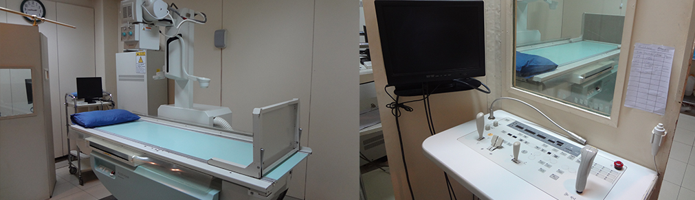
X-Ray
-
COMPUTED RADIOGRAPHY:
We have latest computed radiographic system. Computed radiography (CR) uses very similar equipment to conventional radiography except that in place of a film to create the image, an imaging plate (IP) made of photostimulable phosphor is used. The imaging plate is housed in a special cassette and placed under the body part or object to be examined and the x-ray exposure is made. The imaging plate is run through a special laser scanner, or CR reader, that reads and digitizes the image in computer system. The digital image can then be viewed and enhanced using software that has functions very similar to other conventional digital image-processing software, such as contrast, brightness, filtration and zoom.
-
HYSTEROSALPINGOGRAM:
hysterosalpingogram (HSG) is fluoroscopic X-ray test that looks inside the uterus and fallopian tubes and the area around them. During a hysterosalpingogram, a dye (contrast material) is put through a thin tube that is put through the vagina and into the uterus. It is often done for infertility or any uterine or tubal congenital and acquired pathology.
How To Prepare
- It is done Between 8th – 11th day of menstrual cycle.
- Pregnancy test should be negative.
- Intercourse with the husband is strictly restricted after mensturation and before the examination appointment date.
- Any allergy to iodine dye or any other substance should be informed.
-
BARIUM SWALLOW
A barium swallow is a fluoroscopic test that may be used to determine the cause of painful swallowing, difficulty with swallowing, abdominal pain, bloodstained vomit, or unexplained weight loss.
Barium sulfate is a metallic compound that shows up on X-rays and is used to help see abnormalities in the esophagus. When taking the test, you drink a preparation containing this solution. The X-rays track its path through your digestive system.How To Prepare
- Only 8 hours fasting is required.
-
BARIUM MEAL EXAMINATION
Barium meal examinations are used to study the lower esophagus, stomach and duodenum under fluoroscopic control. This is similar to a barium swallow but its aim is to look for problems in the stomach and duodenum such as ulcers, polyps, tumors, etc. You drink some barium liquid and effervescence, but you then lie on a fluoroscopic table whilst X-ray pictures are taken over your abdomen. It may take a little longer to do than a barium swallow, So that the barium coats all around the lining of the stomach and duodenum.
How To Prepare
- Only 8 hours fasting is required.
-
BARIUM FOLLOW THROUGH
This test is similar to a barium meal but aims to look for problems in the small intestine. Therefore, you drink the barium liquid but then need to wait 10-15 minutes before any X-rays are taken. This allows time for the barium to reach the small intestine. You may then have an X-ray every 30 minutes or so until the barium is seen to have gone through all the small intestine and reached the large intestine (colon). It can be done independently or routinely done along with Barium meal examination.
How To Prepare
- One day preparation is required.
- 4-6 tablets of dulcolax should be taken one day prior to examination.
- 8 hours complete fasting is required.
-
SMALL BOWEL ENEMA (ENTEROCLYSIS)
This test is similar to a barium follow through. However, instead of drinking the barium liquid, a thin tube is passed down your oesophagus, through the stomach and into the first part of the small intestine. Barium liquid is then poured down the tube under fluoroscopic control and spot films taken in multiple intervals.
How To Prepare
- One day preparation is required.
- 4-6 tablets of dulcolax should be taken one day prior to examination.
- 8 hours complete fasting is required.
-
DOUBLE CONTRAST BARIUM ENEMA EXAMINATION
A barium enema is fluoroscopic X-ray examination that can detect changes or abnormalities in the large intestine (colon). An enema is the injection of barium into your rectum through a small tube. In this case, barium coats the lining of the colon. During a barium enema exam, air is also pumped into the colon. The air expands the colon and improves the quality of images. This is called an air-contrast (double-contrast) barium enema.
How To Prepare
- Follow a special diet the day before the exam. You may be asked not to eat meat and hard diet and to drink only clear liquids — such as water, tea or coffee without milk or cream, and clear carbonated beverages.
- Take laxatives daily for 2 days in the night before the examination.
- Fasting is not required.
- Colonic washout will be given in the center 2 hours before the examination.
-
BARIUM ENEMA FOR HIRSCHSPRUNG’S DISEASE
Barium enema examination is also performed for Hirschsprung’s disease in children. No preparation is required for this examination, however 1 -2 days preparation is given to evaluate any other pathology of colon.
-
BARIUM ENEMA FOR POLYP
Barium enema examination is also preform to evaluate polypoidal lesion in colon.
- 2-3 days thorough preparation is given to clean colon from feces which normally obscure small polypoidal lesion.
- No solid diet or semisolid diet or meat/milk containing food is given 2-3 days prior to examination.
- Maximum fluid intake for 2-3 days prior to examination.
-
DISTAL AND PROXIMAL LOOPOGRAM
Distal and proximal loopogram examinations are perform to evaluate leakage, perforation, obstruction of distal or proximal intestine. These are normally performed with high density barium, however it is better to perform it with gastrograffin (water soluble contrast), so that contrast is absorb within 24-48 hours and bowel remain empty at the time of re-anastomosis.
- No preparation is required.
-
DEFECATING PROCTOGRAM
Defecating proctogram examination is perform to evaluate rectal prolapse and other rectal pathology.
- No preparation is required.
-
MAMMOGRAM EXAMINATION
We have dedicated mammographic units in our department and perform mammography examination routinely with internationally recognized protocols. A mammogram can be used either for screening or for diagnostic purposes.
Mammography is a specific type of imaging that uses a low-dose x-ray system to examine breasts. A mammography examination, called a mammogram, is used to aid in the early detection and diagnosis of breast diseases in women as well as in men.
For patients of mammogram, we perform ultrasound examination on complimentary basis to make best diagnosis and correlation between the two modalities, which are beneficial for the patient for correct diagnosis and instant management.-
Avoid deodrants and talcum powder on the day of examination.
Mammogram Recommendations for all women: - Women 40 and older should have mammograms every 1 or 2 years.
- Women who are younger than 40 and have risk factors for breast cancer should ask their healthcare professional whether mammograms are advisable and how often to have them.
- Even women who have no symptoms and no known risks for breast cancer should have regularly scheduled mammograms to help detect potential breast cancer at the earliest possible time.
-
Avoid deodrants and talcum powder on the day of examination.
-
SINOGRAM / FISTULOGRAM EXAMINATION
Fistulograms and sinograms are imaging scans used to assess abnormal tract anywhere in the body. A fistula is an abnormal tract tube connecting two organs, such as the bowel or bladder, or between an organ and the skin. A sinus is an abnormal track or cavity that opens to the skin.
A fistulogram shows a fistula and a sinogram shows a sinus tract. The scans allow doctors to see the connecting tube or track more clearly. It uses iodinated liquid contrast, which shows up well on x-rays.How To Prepare
- Patient should have discharging sinus opening at the time of examination
-
SIALOGRAM
A sialogram is a contrast examination of the salivary ducts and glands.
The salivary glands are located on each side of the face and beneath mandibles. They release saliva into the mouth. It consist of parotid and submandibular sialogram examination.How To Prepare
- No special preparation is necessary before a sialogram. Food and fluid intake do not need to be restricted.
- Bring lemon along with on the day of examination.
-
DACRYOCYSTOGRAM
A dacryocystogram is a special x-ray procedure that is done to visualize the lacrimal duct of the eye following the injection of contrast media into the duct. A fine catheter or special blunt needle is inserted into the tiny duct on the inner corner of the eye and film taken under fluoroscopic control.
How To Prepare
- No special preparation is necessary before the examination.
-
URETHROGRAM
Urethrogram is a routine radiologic procedure used to image the integrity of the urethra and bladder. It consist of ascending (retrograde) and descending (Antegrade) study of urethra.
How To Prepare
- No special preparation is necessary before the examination
-
INTRAVENOUS PYELOGRAM (IVP), IVU AND EU EXAMINATION
An intravenous pyelogram (IVP) is an x-ray examination of the kidneys, ureters and urinary bladder that uses non- iodinated contrast material injected into veins.
When a contrast material is injected into a vein in the patient’s arm, it travels through the blood stream and collects in the kidneys and urinary tract, turning these areas bright white on the x-ray images. An IVP allows the radiologist to view and assess the anatomy and function of the kidneys, ureters and the bladder.How To Prepare
- One day preparation is required.
- 4-6 tablets of dulcolux should be taken one day prior to examination.
- 8 hours complete fasting is required.